Multi-branch Convolutional Neural Network for Multiple Sclerosis Lesion Segmentation
Introduction
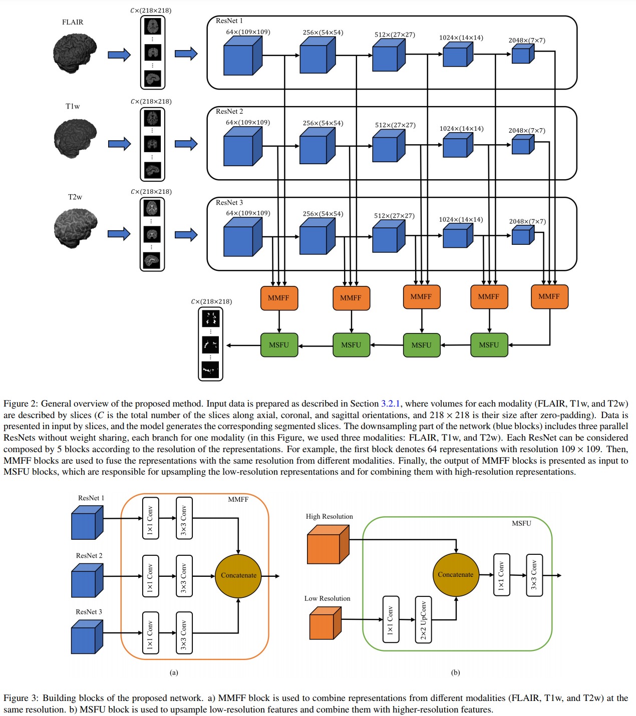
This paper presents what seems to be the state-of-the-art multiple sclerosis segmentation method. This method has three main characteristics:
- A multi-modal ResNet encoder
- A multi-modal fusion at the skip connexion level
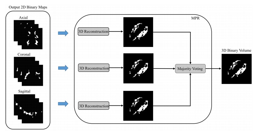
- A majority vote combination of sagittal, coronal and axial prediction.
Proposed method
The proposed network is illustrated in Figure 2. The blue boxes are ResNet blocks, the orange boxes are Multi-Modality Feature Fusion (MMFF) blocks and the green boxes are Multi-Scale Feature Upsampling (MSFU) boxes. The loss implements a soft Dice function:
\[DL = 1-\frac{2\sum_i g_ip_i}{\sum_i g_i^2 \sum_ip_i^2}\]where \(i\) is a pixel index, \(p_i\) is the predicted value for pixel \(i\) (a probability value between 0 and 1) and \(g_i\) is the groundtruth label for pixel \(i\).
While the network processes 2D slices, it produces results for the axial, sagittal and coronal orientations. The three predicted values are then combined via a majority vote (c.f. the following figure )
Results
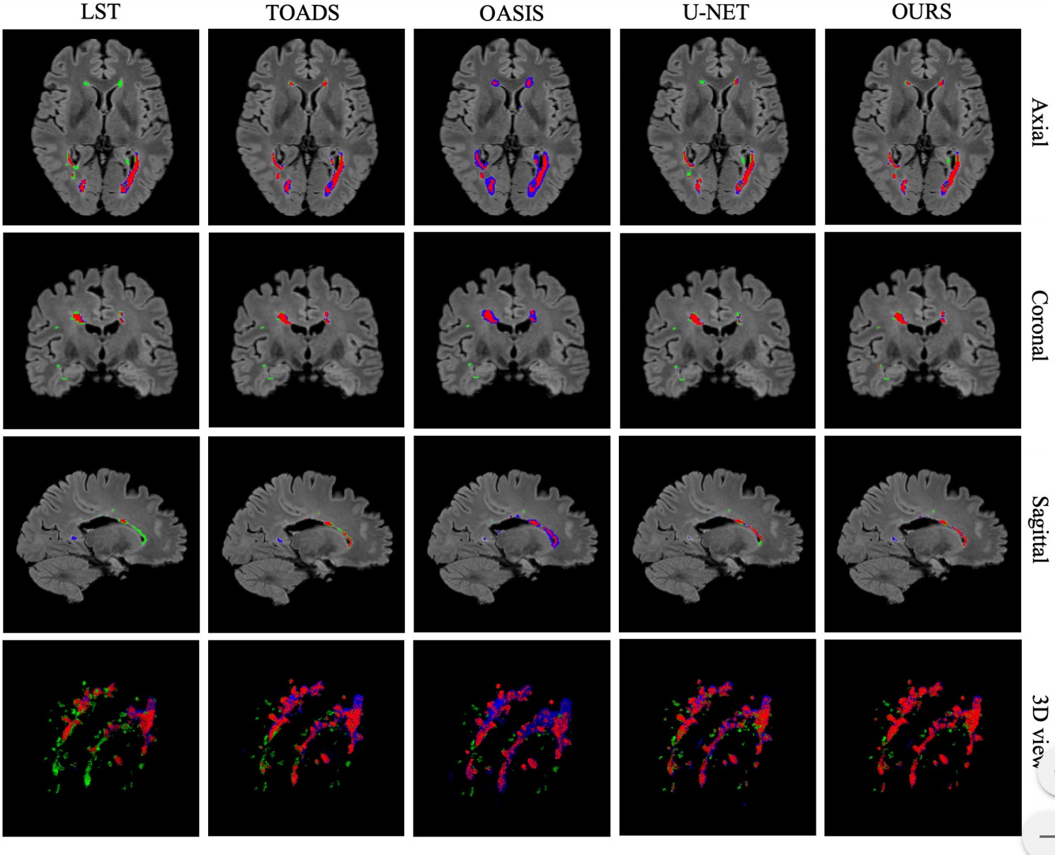
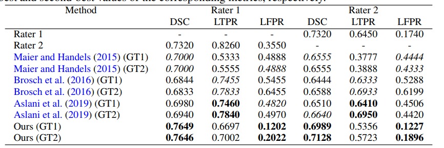
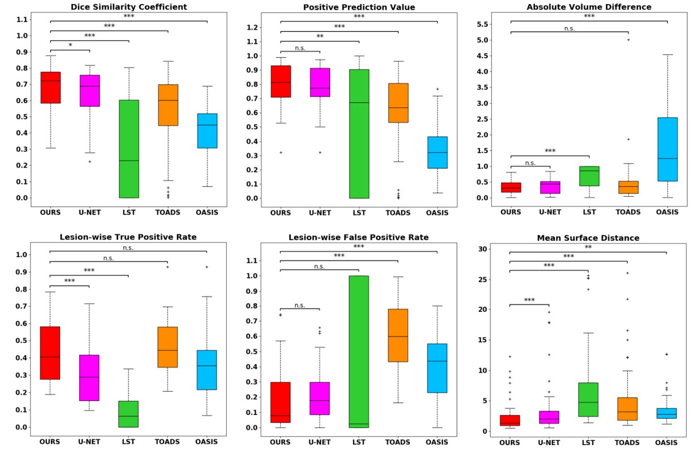
They tested their method on the neuroimaging research unit (NRU) dataset which contains images of 37 patients. While their results are slightly better than those of a UNet, they are getting closer to the inter-observer variation. Unfortunately the paper has no ablation study so it is hard to quantify the benefit of every aspect of the method.
